// Twitter Cards // Prexisting Head The Biologist Is In: February 2018

Monday, February 19, 2018

A Bird's View of Color

Diagram illustrating the frequency sensitivity of three photoreceptors in humans and four photoreceptors in starlings.
Figure from [link].
Most birds have much better color vision than mammals. In general, they have four distinct types of color-sensing cones in their eyes, compared to the usual three for us and two for most other mammals. The fourth cone that birds have is sensitive to ultraviolet (UV), letting them perceive colors we can only imagine. The other three cones don't precisely match up with our three, but they cover basically the same range of frequencies.

To get an idea of what things look like to birds, we have to incorporate that UV information we can't see. Taking photographs of the UV world can take some special equipment, but even consumer grade cameras can be altered to better capture UV light. I've been interested in photography for a while and I've been interested in UV photography, but I haven't yet invested in the equipment I'd need to take UV photos. For now I have to rely on people posting occasional UV photos to get an idea what things look like. (For a good selection of photos in UV and other frequency bands, go take a look at: photographyoftheinvisibleworld.blogspot.com)

You can look at UV light photos next to visual light photos to get an idea of what things look like to birds, but I decided to see if I could do one step better. I wrote a script which takes four image channels (Red, Green, Blue, Ultraviolet) and compresses them into the three we can see (RGB). The math for this is pretty simple and so doesn't match what really happens in detail, but it might help us get an idea of what things look like to birds.

\(R_h = R_b + \frac{G_b}{3}\)
\(G_h = \frac{2(G_b+B_b)}{3}\)
\(B_h = \frac{B_b}{3} + U_b\)

Figure illustrating the four primary colors seen by starlings and the three primary colors seen by humans. Starlings see in the ultraviolet frequency range that humans cannot.
RGBU (bird vision) -> RGB (human vision).
This math is represented visually in the diagram at right. At the top are the four image channels that birds can see and at bottom are the three we can see. All the information that birds can see in their red channel goes into our red channel, along with a third of what birds see in their green channel. The other channels of a bird's vision are similarly apportioned into the channels we can see by moving from top to bottom in the figure.

Conceptually, this is similar to drawing a 3D cube on a 2D sheet of paper. Some information is lost in the transformation, but much of it comes through the process and allows us to visualize something that otherwise can't be done (in 2D). In this case, we're transforming a 4D data structure into a 3D one, that just happens to be presentable as a color photo.


To show what this math means for a photo, I found a nice example paired set of visual and UV images from photographyoftheinvisiblew... to work with.

Single small flower seen in red, green, blue, and UV frequency ranges at top. At bottom-left is an image built from the RGB channels. At bottom-right a version of the image built from RGB and UV channels.
RGBU image channels along top; RGB and compressed RGBU images at bottom.
Original photos from [link].

In UV this flower of the Marsh Marigold (Caltha palustris) is dramatically marked, but in our composite RGBU image it really doesn't stand out that much. Flowers rarely utilize birds for their pollination, so it shouldn't be any surprise that they might not look too dramatic to birds. (Bees can't see red, but can see UV, so they'd have no problem seeing the marks on this flower.)


Can we find some nice UV imagery of something that birds would care about? Well, it's a bit harder to make paired visual and UV photos of a creature which is suspicious of your intentions. Recently, I came across a post by twitter user @JamieDunning illustrating some dramatic fluorescence on the beak of a Puffin specimen he was examining. It occurred to me that something strongly fluorescent should also be UV-dark, since the UV energy is being absorbed and released at visual frequencies instead of reflected. I realized from this I could construct a simulated UV-channel by inverting the fluorescence image (and mapping the images together to correct for the different camera position (and using a bit of artistry to clean up the fluorescence image)). Performing the same image channel compression as I did earlier, we get the lower-right portion of the next figure.

Similar image to above, but the subject is a frozen puffin focused on the beak. In the lower-right image, where a simulated four primary color image is presented with three primary colors, the beak shows a strongly contrasting color pattern not seen in the three color version.
RGBU image channels along top; RGB and compressed RGBU images at bottom.
Original photos from [link].

It would be nice to have a comparable real UV-photo for doing this comparison, but that the simulated bird-vision of the Puffin's beak shows a much greater color contrast than we saw with the Marsh Marigold (and that both results align with the evolutionarily expected results) suggests this might be a useful approach.


My wish-list, money-is-no-option, sort of data for doing this kind of image analysis would be that produced from hyper-spectral imaging. A hyper-spectral camera takes images at a large range of narrow frequency bands. We could map that data (vs. the color sensitivity spectra illustrated in the figure at the top of this post) to what either birds or humans can see, as well as something like the transformation I've described here to illustrate in human vision what a bird could see.

I'm not sure anyone would provide sufficient funding for me to explore this, however.


@JamieDunning recently submitted a paper comparing the original photo with spectrophotometer examination of regions of the bill. I'm looking forward to their paper to see how the results compare to the predictions from my playing around with the math.


References:

Monday, February 12, 2018

Chromosome Painting

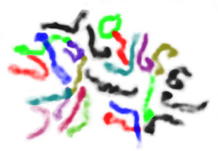
Microscope image of chromosomes, false-colored to help visually distinguish individual chromosomes. The figure at left shows the metaphase chromosomes of a pepper root-tip, in all their squiggly false-color glory. In it you can count the number of chromosomes and (with some little background research) determine the overall ploidy of the source plant. (It has 24 chromosomes, so is a diploid.)

The original image had all the same information, but it was much harder to look at and learn from. This is a fundamental lesson of, and reason for, data visualization.


Microscope image of a cell with condensed chromosomes visible.
Step 0.
The original image comes from Twitter user @ChaoticGenetics. They're studying chile genetics and routinely post cool photos derived from their work. The question paired with this image was, "How many chromosomes does everyone see?" I figured I'd take a stab at it.

Lets dive into the details of how I made my figure. I use GIMP for essentially all my image editing needs. With each step figure I'll include the menu options for each command I use in brackets, so others can repeat the procedure.

0) Load the image with GIMP. Open "Tool Options" [Control-B] and "Layers" [Control-L] windows.

initial image cropped to just show the condensed chromosomes.
Step 1.
White color of the image has been converted to transparency.
Step 3.
1) Select a rectangular region around the interesting looking chromosomes, then crop [Image > Crop to Selection] the image.

Background of the condensed chromosomes has been erased.
Step 2.
2) Select the "Eraser Tool" and erase all the background color and spots that don't appear as chromosomes.

3) Right-click on the image in the layer window. Select, "Add Alpha Channel". Discard the color information in the image [Colors > Desaturate]. Remove the background color [Colors > Color to Alpha (Set "From:" color to white.)].

White background is restored to the image.
Step 4.
4) From the layer window, make a new image layer filled in white. Move this layer beneath the image layer. Select the image layer.

A single condensed chromosome has been false-colored with purple.
Step 5.
5) Using the "Free Select Tool", draw around a visually distinct chromosome. Invert the color of the selection [Colors > Invert]. Change the color of the selection [Colors > Components > Channel Mixer... (red=50,0,0; green=0,0,0; blue=0,0,50)].

A second condensed chromosome is false-colored, this time in green.
Step 6.
6) Many of the chromosomes in this example are adjacent or overlapping with another. For these, we have to use some knowledge about chromosomes and some artistry. Lets have a look at the cluster here highlighted in green.

Green false-colored chromosome broken up into three parts, each colored part colored differently (blue, red, and green).
Step 7.
7) At this scale, chromosomes are essentially linear structures. They don't branch and they don't loop. From this we can tell the green feature in step 6 is actually three chromosomes. I cut each chromosome out of the image and pasted into a new layer. From there I could clean up their shape a little before changing the colors and recombining them.
Several chromosomes have been highlighted in various colors. Large aggregate of condensed chromosomes that can't be visually separated is colored in pink.
Step 8.

8) Going progressively through the image, isolating and coloring the most apparent chromosomes at each stage, we come to 16 chromosomes that we can be confident about. (So, our cell isn't a haploid with 12 chromosomes.)

We're left with the region at left I've highlighted in pink. This region would need to account for a further 8 chromosomes to reach the expected diploid count of 24 in total. Though there are probably a few chromosomes in this region that we can confidently separate, much of it is down to guesswork.

It is possible for this specific pepper plant to have fewer chromosomes. Though it is unlikely for a chromosome pair to be lost, since each has been conserved over a long time period and likely contains critical genes, it is common enough evolutionarily for chromosomes to fuse. That pink mess could hypothetically be 6 or 4 chromosomes, though this one image isn't sufficient evidence to make me think it is likely. If the same pattern is shown in a few more images from the same plant, especially if the chromosomes are better spread, then I'd start to consider that as increasingly likely.


For now, the balance of the evidence leads me to think there are 24 chromosomes and they're just not perfectly isolated. So, I divided the uncertain pile of chromosomes into the number that I expect are remaining. Any figure you make will invariably include your assumptions. The key is to try and make those assumptions reasonable or at least apparent to the reader (though this may require some nice caption-writing).

Interestingly, there's a protocol which can experimentally produce the sorts of painted chromosomes we're simulating here. Fluorescent In-Situ Hybridization (FISH) relies on making DNA probes which are stained a unique color for each chromosome. When the probes are applied to a chromosome spread, the result helps visualize chromosome crossovers, deletions, and other large scale alterations that can be important in diagnosing cancer and other disorders. The setup work for this is pretty intense, so it's probably not going to be used for the simple task of seeing how many chromosomes a plant has.


While I was in grad school, I routinely modified figures from papers I was reviewing for in-class (or in-lab) presentations. Usually highlighting different components of the figure in different colors (like here), to make them stand out more when displayed. I was doing the hard work of figuring out the important parts of the figures so students watching my presentation didn't have to. My goal was for them to focus on what I was saying about the figures and see what wanted them to see at a glance.

Using colors to present different partitions of a larger dataset ended up being central to my last large graduate project (YMAP) as well as an important part of my current [non-academic] job. While using colors for data presentation, it is important to keep in mind that not everyone has the same ability to see color. The most common forms of color-blindness are often called Red-Green-colorblindness. From this, it is a good idea to try and avoid the commonly used Red-Green color scheme seen so often in biology research figures. (Blue-Yellow is a good alternative, but there are subtleties I'll have to go into later.) Being conscious of the issues means they will inform your decisions, even if you're not fully aware of the topic.


This post was inspired by a conversation over on Twitter. (You can follow me there as @thebiologistisn.)

The original picture of the chromosome spread was made by @ChaoticGenetics, who gave permission for me to use it in this post.


References:
https://twitter.com/ChaoticGenetics/status/9602424397463060

Monday, February 5, 2018

Speculative Biology: 3-Way Reproduction

The other day I found myself thinking about what would be the fundamental biological characteristics of a species having a system which depended on three individuals, instead of the two or one we're used to, for each reproduction event. (This is a distinctly different concept from a species having multiple sexes or genders. See references at end for examples of these.)

To simplify the discussion, I'm going to start with a big assumption that the hypothetical organism only differs from what we see here in that it has a 3-way reproduction system. It is carbon based, it has DNA organized into chromosomes, etc. Breaching that huge assumption would introduce far more variables into the discussion, when I'm only interested in the basics of sexual reproduction for this discussion.


There are different ways for Earth biology to control the sex of individuals. Sex in some species is driven by genetic differences (mammals and birds). In some it is driven by the number of chromosomes (bees and wasps). In some it is driven by temperature differences (reptiles and amphibians). In others the sex changes with age or social situations (some fishes). In large groups, there are almost always exceptions to the general patterns.

Almost all of these cases involve some chromosomes being contributed to an offspring by both a female and a male. Haplodiploidy in bees/wasps is an interesting exception. (Males grow from unfertilized eggs, while females grow from fertilized eggs.)


For our 3-way reproduction discussion, lets start by assuming each of the three individuals contributes chromosomes equally to each offspring. (Later we'll examine a more complicated case.)

We can easily enough abstract the concept of a Punnett square into higher dimensions, though it does get difficult to simply convey the results in a 2D format. In the regular version, the possibilities for a single chromosome contributed from each parent are aligned along the top and side edge of the table. For a 3D version we'll do the same, but split the contributions from the third parent into three sub-tables (left to right) with the third parent contribution at the upper-left corner of each. (I've added some color highlights to help visualize the contributing parent for each chromosome as well as the sexes of the potential offspring.)

Punnett square (and "cube") for 2-way and 3-way crosses.


The first observation that stands out from this is the difference in predicted sex ratios of the offspring. In our 2-way system, the calculations implies a 1:1 ratio. In the 3-way system, the calculations implies a 1:2.5:1 ratio between the three sexes of offspring.


Next lets discuss something analogous to the haplodiploidy of bees/wasps, where males only contribute chromosomes to their daughters.

Punnett square (and "cube") for 2-way and 3-way crosses, with haplodiploidy.

This shows the same possibilities for sex ratios of offspring. Since we're using bees as a model here, it's a good time to introduce the idea that a creature doesn't have to produce offspring at the ratios suggested by such simple calculations. Bees produce very few males, and only when needed for fertilization of new queens. Similarly, a hypothetical 3-way reproducing species could easily adjust the sex ratio of its offspring to be different from what the above calculations suggest.

An abstraction from Fisher's Principle (http://the-biologist-is-in.blogspot.com/2015/12/evolutionary-battle-of-sexes.html) suggests most species would evolve towards a 1:1:1 ratio between the three sexes. Cases where this wasn't the case would be interesting.


I imagine one sex evolving into an approximation of female (with a large immobile gamete), while the other two sexes evolving into an approximation of male (with smaller, more mobile gametes). It gets much more difficult to make predictions beyond this point, though a couple examples inspired from fiction and biology come to mind.

Maybe the two male-equivalents would actively court each other and then seek out the a female-equivalent together as a pair. This seems to be the pattern described for the fictional Pierson's Puppeteers (though their sexual biology is rarely detailed in the author's stories about them).

Maybe the male-equivalents would independently seek out the female-equivalent. I imagine something similar to the Deep-sea Anglerfish, with smaller male-equivalent individuals fusing to a larger female-equivalent and waiting for the opportunity to contribute to offspring when all three sexes have joined the party.


In the grand scheme of things, I expect biological systems requiring three individuals for reproduction would be rare in the cosmos. At an early evolutionary stage, any organisms which only required two partners to reproduce would probably out-compete those requiring three partners simply because it would be easier to arrange appropriate matings. This isn't to say it wouldn't happen, since all sorts of strange things happen in biology.

The Red Queen hypothesis (http://the-biologist-is-in.blogspot.com/2014/04/oxalis-and-red-queen.html) suggests why larger species don't simply have one sex. Yet, we do see this from time to time.


References