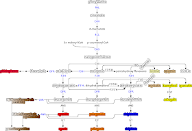
The carotenoid pigment pathway I discussed in those earlier articles was relatively simple. A single main pathway, with a couple branches. The anthocyanin pathway figure at above-right is a bit more complicated. The figure is a consensus pathway, built from research in a few different species. There are definitely more pieces that could be added, but this amount is a good start. The colored highlights are intended to represent the colors of those chemicals. The lower red, orange, and blue pigments are anthocyanins, the pigments responsible for the color of many flowers (and other plant parts). The white-to-brown gradient highlight is for the proanthocyanidins. They oxidize over time, changing from clear to brown. The red pigment at upper-left is found in some trees, but I wasn't able to find too much information about them. The yellow pigments at right are found in various plants and plant parts, but they're not generally the source for bright yellows in flowers. (The enzyme FGT leading to astragalin at far right is something I made up, since I couldn't find any research naming the enzyme performing that step.)
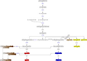
At left is a heavily reduced version of the first figure, trimmed to an approximation of what seems to be going on in common beans (Phaseolus vulgaris). Combinations of the yellow, red, blue, and brown pigments seem to be responsible for most of the variations in color that we see in dry beans. I've seen some evidence for a brown pigment derived from the yellow ones here, but I haven't found any research clarifying the chemistry involved. There's the possibility of some green pigments made up from a different metabolic pathway, but I haven't found sufficient research about them to know if they're represented in beans.
Various of the trimmed compounds are also found in common beans, but they don't seem to be found in significant amounts. The orange pelargonidin pigments have been reported in some bean varieties, but I've never come across a common bean that has a color dominated by orange pigment. There might be orange examples from P. coccineus, the scarlet runner bean, but I'm still investigating this.
Various of the trimmed compounds are also found in common beans, but they don't seem to be found in significant amounts. The orange pelargonidin pigments have been reported in some bean varieties, but I've never come across a common bean that has a color dominated by orange pigment. There might be orange examples from P. coccineus, the scarlet runner bean, but I'm still investigating this.
The colors of beans drew attention far before we had any understanding of the physiology of the pigments involved. Much of the early published research into bean colors sought to identify different genes responsible for the traits. Eventually the gene labels assigned by different authors got correlated with each other and the set of labels for important color genes became standardized. Even more recently, there have been efforts to identify the molecular mechanism behind the different classical gene labels. Some gene labels are now associated with specific enzymes or other genes important in the flavonoid pathway.
- R [red] : Enzyme F3'H, or more likely a transcription factor driving F3'H in the seed coat. F3'H is important for stress response in plant tissues and so is unlikely to be absent even when the enzyme isn't active in the pathway.
- V [violet] : Enzyme F3'5'H. This one isn't as important as F3'H and is entirely absent in many plants.
- J : Pretty solidly identified as the enzyme DFR.
- P : A transcription factor driving expression of several genes important in the flavonoid pathway. In the figures above, the regulated enzyme targets are drawn in blue.
- B : A transcription factor driving expression of chalcone synthase (CHS) and/or chalcone isomerase (CHI).
- G : A transcription factor leading to increased levels of astragalin, perhaps by driving expression of FLS and/or FGT. Likely has other impacts, but I haven't found sufficient research.
Tracking down which gene was associated with which step in the pathway was tricky. Many of the older papers had models for what a given gene did, but then those models were overturned by more recent research. The paper identifying V as being the gene for the enzyme F3'5'H was only published in March 2022. Finding that paper got me interesting in trying to see how many of the others could also be associated with a specific part of the pathway. The other gene notes above came from the scattered papers linked in the references section, though few were specifically the point of the papers.
My goal was to better understand what the gene labels were doing, so I could better figure out what genes were likely to be involved in the beans I was growing and crossing. I'll write more on that another time.
References
- Related blog posts:
- https://the-biologist-is-in.blogspot.com/2014/04/the-color-of-tomatoes.html
- Carotenoid pigments in tomatoes.
- https://the-biologist-is-in.blogspot.com/2015/11/the-color-of-peppers-2.html
- Carotenoid pigments in peppers.
- https://the-biologist-is-in.blogspot.com/2018/10/the-color-of-beans-1.html
- Introduction of my #BlueBeanProject.
- https://the-biologist-is-in.blogspot.com/2022/12/the-color-of-beans-2.html
- Status update of my #BlueBeanProject.
- https://the-biologist-is-in.blogspot.com/2019/11/biology-of-blue.html
- Discussions around the chemistry of blue in biology.
- Papers related to anthocyanin pathway in bean, cotton, etc:
- http://arsftfbean.uprm.edu/bic/wp-content/uploads/2018/04/ChemistrySeedCoatColor.pdf
- https://nph.onlinelibrary.wiley.com/doi/full/10.1002/ppp3.10132
- https://www.ncbi.nlm.nih.gov/pmc/articles/PMC3602603/
- https://link.springer.com/article/10.1007/s11738-011-0858-x
- https://pubmed.ncbi.nlm.nih.gov/28981784/
- https://bmcplantbiol.biomedcentral.com/articles/10.1186/s12870-019-2065-7
- https://pubmed.ncbi.nlm.nih.gov/35289870/
- https://squashpractice.com/2011/10/08/bean-genes/
- https://journals.ashs.org/jashs/view/journals/jashs/124/5/article-p514.xml
- https://www.frontiersin.org/articles/10.3389/fpls.2022.869582/full#ref33
- https://journals.ashs.org/downloadpdf/journals/jashs/120/6/article-p896.pdf
- https://journals.ashs.org/downloadpdf/journals/jashs/125/1/article-p52.pdf
- https://www.semanticscholar.org/paper/Allelism-Found-between-Two-Common-Bean-Genes%2C-Hilum-Bassett-Shearon/f9cef3175289b7d2822461b9d495d8885bb67a48
- https://www.semanticscholar.org/paper/Inheritance-of-Reverse-Margo-Seedcoat-Pattern-and-J-Bassett-Lee/7557538290b700d1fd980a24fba3148846861690
- https://www.semanticscholar.org/paper/The-Margo-%28mar%29-Seedcoat-Color-Gene-Is-a-Synonym-%28-Bassett/d1c58ec1fa0bf9e500d8bd48364a61568b0b7a11
- https://naldc.nal.usda.gov/catalog/IND92036951


No comments:
Post a Comment