// Twitter Cards // Prexisting Head The Biologist Is In: color
Showing posts with label color. Show all posts

Tuesday, August 29, 2023

The Color of Beans 6

Cluster of bright red bean flowers.My ongoing project to produce distinctly blue dry beans occasionally throws a surprise at me. This spring, two plants with bright red flowers appeared in one of my gardens. The blue lines had until now showed a mix of white and pale pink flowers, so this change was rather dramatic.

I immediately began thinking about how these plants came to be. They clearly grew from blue seeds I'd planted, but that was all I knew initially. Last year I had some (more or less) intentional hybrids turn up in my blue bush bean patch, so I thought they might have represented back crosses to the parental blue line.

In another garden this year is filled with an F2 population from that intentional hybrid I mentioned. If the red flower color came from a back cross, then the same color should also turn up among the F2 plants. Only white and pale pink flowers were evident when they started blooming, thus the red flower did not come from a back cross.

I did have one red-flowered Phaseolus coccineus (var "Insuk's Wang Kong") growing last year. The seed germinated late and the vine never prospered. It had only one small flower cluster and produced a single pod with a single seed. It was also on the far end of the garden from the blue bush population, with rows of large tomato and other bean plants in between. I didn't expect there would be any chance for pollen transfer like this, even though I was hoping there might be some crosses with more nearby plants because I'd been wanting to work with a P. vulgaris x P. coccineus hybrid for years.

Dry beans. Most are tan with dark brown streaks, but a few are solid blue.

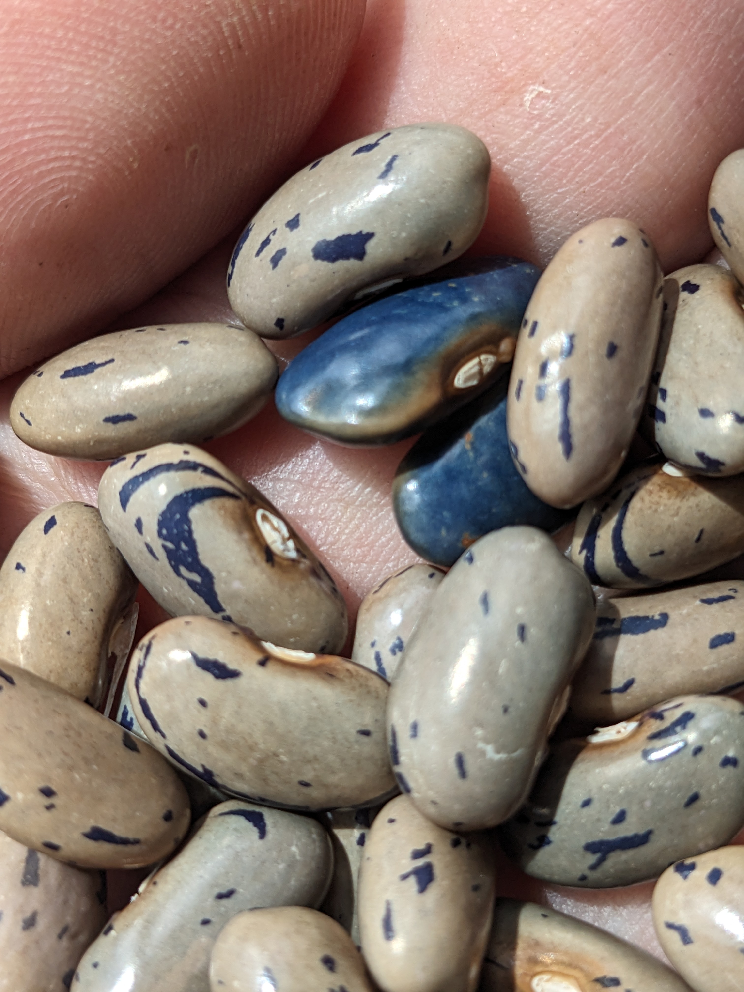
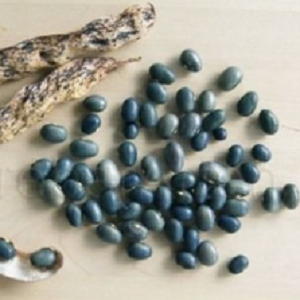
I was eagerly waiting for the first seed pods from the red flowered plants to mature, as their seed color would likely be the definitive evidence for the plants being hybrids. The first pods broke open to reveal tan seeds marked with dark blue. The speckled trait came from the P. coccineus parent and the blue color came from the P. vulgaris parent (the blue seeds here for comparison purposes).

Hybrids between P. vulgaris and P. coccinues are often described as having growth issues or have poor seed-set, though the literature and experience of others seems to be quite variable. Fortunately, my hybrids show no such issues but perhaps they may show up in the next generations.


Because the P. coccineus parent's seeds were a pale purple, I was expecting the hybrid to turn up with a distinctly purple color and I wouldn't see the blue again until the next generation. Since the speckled trait seems to be dominant, I would expect 75% of the next generation to also show speckles. I can't really predict what the colors in the next generation will be, since I don't have a good idea of what the mixed up genetics will do.

I have read from a few references that the P. coccineus chromosomes are preferentially lost vs the P. vulgaris chromosomes in subsequent generations, so any predictable genetics ratios are likely to be distorted significantly. I'll just have to find out. Because of this tendency, similar crosses have been used before to introgress traits into P. vulgaris, such as disease resistance factors. I'm hoping I can stabilize the flower color over the next few years, as well as the tendency to bloom very well.

References:

  1. Interspecific hybridization between cultivated american species of the genus Phaseolus.
  2. Embryo development in reciprocal cross of Phaseolus vulgaris L. and P. coccineus Lam.

Friday, February 24, 2023

The Color of Beans 5

Pile of blue dry beans.

After several years of looking for a truly blue dry bean, I started actively working to produce my own variety in 2018 when I was gifted a few bluish-black hybrid seeds from a bean collector. At the end of 2022, I was essentially done. Five years of selecting the best blue seeds each season and my new variety finally produced a consistently blue crop.

There's probably still going to be a few more years of increasing the seed before I'm prepared to distribute them in some fashion. Along the way I'll be selecting for increased productivity and having to figure out where I can grow more of them each year.

Anthocyanin pathway diagram, emphasizing red and blue anthocyanins, as well as yellow astragalin.

Since harvesting the most recent crop, I've been digging into what research literature there is about the genetics and biology of color in beans. This was initially just to get a better idea of what was going on in my beans. Along the way I realized I could probably use the knowledge I had gained to intentionally make new blue varieties.


The initial random hybrid was between a black variety (Haudenosaunee Skunk) and a yellow-tan variety (Arikara Yellow). That color combination would clearly work as parents in a new cross, but I realized there was a combination of parental color genetics that would probably make it easier to recover the right combination of genes to produce a blue color.

The color of black beans is caused by a combination of red, blue, and yellow-brown pigments at high intensity. In Phaseolus vulgaris, a small number of genes lead to this color: R ("red"; likely a transcription factor driving enzyme F3'H), V ("violet"; enzyme F3'5'H), and then B ("brown"; transcription factors enhancing CHS/CHI, darkening all downstream pigments). A yellow-brown bean is caused by all three genes being inactive (written as "rvb"). There are a few other genes that seem to impact exactly what shade of brown color may result, but there doesn't seem to be as much clarity in the research literature about that part of the pigment pathway.

The difference between a purple and a black bean is that the purple bean has an inactive version of B (b). Crossing a purple bean (RVb) to a yellow bean (rvb), where both have the recessive inactive b allele, reduces the genetic possibilities in the F2s to only those impacting the red and blue pigments. Among the F2 plants, we should see the ratios of a dihybrid cross play out. 9 purples, 3 reds, 3 blues, and 1 yellow-brown. 3/16 isn't that bad and I can easily grow enough plants to expect to be able to find the blues I'm looking for.

At left, purple dry beans. At right, yellow dry beans.
To test this model I've been building, I decided maybe I could try to make a blue version of one of the other domesticated bean species. Most P. coccineus (runner or ayacote) beans I'd ever seen before were lilac and heavily spotted with black. After digging around for a while, I eventually ordered some mixed color packages from some Mexican bean importers in the south-west. Among those, I was able to select out seeds with the colors I was looking for.

While I was waiting for those to arrive, I was also looking for P. lunatus (lima) beans with the same colors. The yellow-brown variety I found is called Pima Orange. I ordered the purple variety from someone on Ebay and am still waiting for them to arrive.

I wasn't able to find any sign of a purple version of P. acutifolius (tepary) beans. I did find black and yellow versions that I could use with more difficulty. I'll keep looking for a purple version and may eventually decide to try using a black variety instead, but for now I'll hold off on trying to make a blue variety of this species.


The color of a seed is determined by the genes of the mother plant, so it can take some tricks to sort things out. The plan will take a few years to play out.

  • 2023 : Plant purple & yellow seeds on a common trellis. Some harvested seeds may be hybrids, but we can't identify them yet. Save them separated by color.
  • 2024 : Plant yellow seeds only. Save harvested seeds by color.
    • Yellow seeds : mother plant wasn't hybrid.
    • Purple seeds : mother plant was a hybrid between purple & yellow plants. These seeds will grow into F2 plants.
  • 2025 : Plant purple seeds only. Save harvested seeds by color.
    • The F2 plants should fall into four categories (9 purple : 3 red : 3 blue : 1 yellow). I've already determined I can grow F2 plants in a mass planting and observe the expected ratios in the cumulative produced seed counts.

P. coccineus and P. lunatus are strong out-crossers, so I can rely on bees and other pollinators to do the work of transferring pollen for me. The two species can't cross, so I can do both parallel experiments in a relatively small garden space. To increase the odds of the yellow seeds produced in 2023 being hybrids, I can plant many more purple seeds than yellow (or even only one yellow seed among many purple). This will result in most flowers that a yellow-seed plant can cross with being those from purple-seed plants.

If no purple seeds turn up in 2024, then no useful cross-pollinations happened in 2023. In 2025 I would then plant purple and yellow seeds to try and find crosses again. There may be some hybrid seeds among the purples, but distinguishing them from the non-hybrids would take a couple more years and require individual plants to be grown on separate trellises. That's more work than I want to put into it, hence designing the plan the way I have.


This plan assumes the genes driving F3'H (red anthocyanins) and F3'5'H (blue anthocyanins) are unlinked in these species. I know in P. vulgaris the two genes are not tightly linked. If they were, I would not expect to have been able to find the initial blue-seeded plant so easily as I did. That it is possible to make hybrids between P. vulgaris and P. coccineus tells us their genomes are organized in largely the same fashion, so the two genes should be similarly not tightly linked in P. coccineus. Making hybrids with P. lunatus is harder, but still possible, so similarly I don't expect the two genes to be tightly linked.

Why aren't there already blue varieties of these species available? I don't know, but I feel it might be the same reason that blue varieties of P. vulgaris are so very rare, whatever that is. That and the vast majority of people who have ever grown beans have not been geneticists backed up with decades of published research into the biology of bean pigments. In a few years, I hope to have remedied this absence.


References:
  1. Bean varieties:
    1. Haudenosaunee Skunk: https://exchange.seedsavers.org/page/variety/id/193592
    2. Arikara Yellow: https://www.seedsavers.org/arikara-yellow-bean
    3. Pima Orange : https://www.nativeseeds.org/products/pl011
    4. Black Tepary : https://www.nativeseeds.org/collections/tepary-beans/products/pt082
    5. S'oam Baw Tepary : https://www.nativeseeds.org/collections/tepary-beans/products/pt120
  2. Blog posts:
    1. https://the-biologist-is-in.blogspot.com/2018/10/the-color-of-beans-1.html
    2. https://the-biologist-is-in.blogspot.com/2022/12/the-color-of-beans-2.html
    3. https://the-biologist-is-in.blogspot.com/2023/01/the-color-of-beans-3.html
    4. https://the-biologist-is-in.blogspot.com/2023/02/the-color-of-beans-4.html
  3. Bean species hybrds:
    1. https://link.springer.com/article/10.1007/BF01902923
    2. https://www.semanticscholar.org/paper/Hybrid-plant-of-Phaseolus-vulgaris-L.-and-P.-L.-by-Kuboyama-Shintaku/c20048408e38632b8ae8fe45d234e967ad57df2d

Friday, February 10, 2023

The Color of Beans 4

In my last post (https://the-biologist-is-in.blogspot.com/2023/01/the-color-of-beans-3.html), I shared a couple figures illustrating the flavonoid/anthocyanin pigment pathway in plants and in common beans (Phaseolus vulgaris) specifically. A couple days later, I found some additional evidence which led me to feel the need to update my figures somewhat.


Starting section from the top: phenylalanine to cinnaminate to 4-coumerate to p-coumaroyl-CoA (+ 3x malonyl-CoA) to naringen chalcone to naringen. Naringen goes left and right to eriodictyol and pentahydroxy flavone. Eriodictyol goes left to flavan-4-ols and then to phlobaphenes (highlighted in red). Eriodictyol goes right to tricetin. Naringen goes right to apigentin (highlighte light brown). Pentahydroxy falvanone goes right to luteolin (highlighted pale yellow). Naringenin goes down to dihydrokaempferol. Eriodictyol and dihydrokaempferol go left to dihydroquercetin. Pentahydroxy flavanone and dihydrokaempferol go right to dihydromyricetin. Dihydroquercetin goes right to quercetin (highlighted in yellow). Dihydrokaempferol goes right to kaempferol (highlighted in yellow) and then down to astragalin (highlighted in yellow). Kaempferol goes to a series of question marks highlighted in a gradient from white to brown. Dihydromyricetin goes right to myricetin (highlighted light brown). Dihydroquercetin goes down to leuocyanidin. Dihydrokaempferol goes down to leucopelargonidin. Dihydromyricetin goes down to leucodelphinidin. Leucocyanidin goes down to cyanidin and then cyanin (both highlighted red). Leucopelargonidin goes down to pelargonidin and pelargonin (both highlighted orange). Leucodelphinidin goes down to delphinidin and delphinin (both highlighted blue). Leucocyanidin, leucopelargonidin, and leucodelphinidin go left to 2,3-trans-flaven-3-ols (catechin) (highlighted in in a gradient from white to brown). Cyanidin, pelargonidin, and delphinidin go left to 2,3-cis-flaven-3-ols (epecatechin) (highlighted in a gradient from white to brown). Catechin and epicatechin go down to proanthocyanidins (highlighted in a gradient from white to brown). Luteolin, apigenin, and tricetin have a group label 'flavones'. Myrictein, kaempferol, and quercetin have a group label 'flavonols'.  The figure has enzyme labels at most steps.  In the top starting section: PAL, C4H, 4CL, CHS, and CHI. Naringenin to eriodictyol is F3'H. Naringenin to pentahydroxy flavanone is F3'5'H. Eriodictyol, naringenin, and pentahydroxy flavanone to tricetin, apigenin, and luteolin are FNS. Eriodictyol, naringenin, and pentahydroxy flavanone to to dihydroquercetin, dihydrokaempferol, and dihydromyrecetin are F3'H. Eriodictyol to flavan-4-ols is DFR. Dihydrokaempferol to dihydroquercetin is F3'H. Dihydrokaemperol to dihydromyricetin is F3'5'H. Dihydroquercetin, dihydrokaempferol, and dihydromyricetin to quercetin, kaempferol, and myricetin are FLS. Dihydroquercetin, dihydrokaempferol, and dihydromyricetin to leucocyanidin, leucopelargonidin, and leucodelphindin are DFR. Leucocyanidin, leucopelargonidin, and leucodelphindin to cyanidin, pelargonidin, and delphinidin are ANS. Cyanidin, pelargonidin, and delphinidin to cyanin, pelargonin, and delphinin are GT. Leucocyanidin, leucopelargonidin, and leucodelphindin to catechin are LAR. Cyanidin, pelargonidin, and delphinidin to epicatechin are ANR.
Certain combinations of published genes lead to production of a brown pigment when an excess of yellow astragalin would be expected. Here I've made up an enzyme called FNR (FlavoNol Reductase) leading to production of the pigment, modeled after the production of proanthocyanidins by ANR (Anthocyanin Reductase).

The evidence for a brown pigment derived from the yellow pigment pathway comes from the gene B changing a bean color from yellow-brown to mineral/dark-brown when present. B is thought to be a transcription factor that enhances the expression of other pathway genes. Yellow can only arise when red and blue pigment branches are absent, so the brown pigment produced with B can't be the brown proanthocyanidins derived from the red and blue pigment branches. Instead it must be an analogous pigment produced from kaempferol and/or astragalin. I haven't found any research papers discussing this pigment pathway branch, but all the evidence seems to point to it being there anyhow.

This version of the flavonoid pigment pathway is trimmed to be limited to the main pigments (cyanidin, pelargonidin, delphinidin, & astragalin) along with the core of the pathway leading to them and the metabolites derived from them.
The figure trimmed to the metabolites and pigments significant in common beans... now includes pelargonidins. This version of the figure has different thickness arrows in places to illustrate when one branch has a higher priority, when one branch of metabolites is more likely than another.

Dihydrokaempferol is a central metabolite for all the different pigment branches in the pathway. F3'H and F3'5'H leading to red and blue pigments have the strongest branches, followed by FLS leading to yellow pigments, and then last is DFR leading to orange pigments. For the orange pigments to dominate the final color, it looks like the other three branches have to be disabled or significantly reduced.

pale tan beans with orange-toned eyes.
So. Are there orange varieties of common bean? Maybe. Instagram user @g3netic_lottery shared a mixed variety of beans they grew this year and some of them had a distinctly orange-ish color, especially in the hilium ring. Now, I'm not entirely sure that is the color of pelargonidin, but it is the closest to orange I've seen in common beans. It's enough of a suggestion that I decided to rework the pathway figure to include it for this species.

I still think it would be really cool to find (or breed up) a variety that had distinctly orange color all over the seed. These beans at right hint that it is feasible if one has the right sort of luck. @g3netic_lottery on Instagram grows beans and other crops in South Africa. I'll be keeping my eye out for useful seeds more accessible to me that might help me get to a nicely orange common bean.


The other domesticated bean species will have very similar pathways as the common bean, but the specific available mutations are going to be different. Maybe we can find a good pelargonidin orange color in one of them. I spent some time looking around for orange seeded varieties of the other species, with limited luck.

  • Runner beans (P. coccineus): This photo of "Ayacote Mexican" beans includes some very orange looking seeds, but I have no idea how much that reflects reality.
  • Lima beans (P. lunatus): "Pima Orange" has seeds that look yellow-brown or orange depending on the photo. Again, it is hard to say what they really look like.
  • Tepary beans (P. acutifolius) : There are some photos around of very orange tepary beans, but I can't find any varieties available for sale that look anything near orange.
  • Year-long beans (P. dumosus) : This species is interesting, but only grown in a limited area. I found several articles about it, but I have yet to find anyone selling seeds.
Mix of dark purple and black beans.
I ordered the runner and lima bean varieties above, so I can see what their colors actually look like. It is so very easy to intentionally or accidentally tweak the color of a photo, so the color of photos online isn't always something you should trust.

The runner beans turned up first. Unfortunately, there was nothing like orange among them. I did get the lovely dark purple beans at left though, which are perfect for another project I'll describe in another post. After further looking around, I found a different vendor selling ayacote beans that are at least advertised with some nice orange tones. As they too are selling the beans intended for food use, there's no guarantee any yellow seeds will turn up.

Mix of orange colored beans with variable dark markings.
The lima beans arrived a few days later. When I opened the package, I was greeted by seeds with a surprisingly nice orange color! Now, this isn't anything like the orange you get from beta-carotene in some carrots and tomatoes, but this might just be what a pelargonidin orange looks like.

Alternately, this could be a mix of a yellow and a brown pigment. There may be some simple basement-lab tests I can do to help me identify the pigment, but that will take some further research.

In either case, I think it is entirely reasonable to expect a similar orange colored common bean should be possible. It's just a matter of finding the right mutations and crossing them into the same plant.

References:

Friday, January 20, 2023

The Color of Beans 3

Diagram illustrating the flavone pigment pathway.

Starting section from the top: phenylalanine to cinnaminate to 4-coumerate to p-coumaroyl-CoA (+ 3x malonyl-CoA) to naringen chalcone to naringen. Naringen goes left and right to eriodictyol and pentahydroxy flavone. Eriodictyol goes left to flavan-4-ols and then to phlobaphenes (highlighted in red). Eriodictyol goes right to tricetin. Naringen goes right to apigentin (highlighte light brown). Pentahydroxy falvanone goes right to luteolin (highlighted pale yellow). Naringenin goes down to dihydrokaempferol. Eriodictyol and dihydrokaempferol go left to dihydroquercetin. Pentahydroxy flavanone and dihydrokaempferol go right to dihydromyricetin. Dihydroquercetin goes right to quercetin (highlighted in yellow). Dihydrokaempferol goes right to kaempferol (highlighted in yellow) and then down to atragalin (highlighted in yellow). Dihydromyricetin goes right to myricetin (highlighted light brown). Dihydroquercetin goes down to leuocyanidin. Dihydrokaempferol goes down to leucopelargonidin. Dihydromyricetin goes down to leucodelphinidin. Leucocyanidin goes down to cyanidin and then cyanin (both highlighted red). Leucopelargonidin goes down to pelargonidin and pelargonin (both highlighted orange). Leucodelphinidin goes down to delphinidin and delphinin (both highlighted blue). Leucocyanidin, leucopelargonidin, and leucodelphinidin go left to 2,3-trans-flaven-3-ols (catechin) (highlighted in in a gradient from white to brown). Cyanidin, pelargonidin, and delphinidin go left to 2,3-cis-flaven-3-ols (epecatechin) (highlighted in a gradient from white to brown). Catechin and epicatechin go down to proanthocyanidins (highlighted in a gradient from white to brown). Luteolin, apigenin, and tricetin have a group label 'flavones'. Myrictein, kaempferol, and quercetin have a group label 'flavonols'.

The figure has enzyme labels at most steps.

In the top starting section: PAL, C4H, 4CL, CHS, and CHI. Naringenin to eriodictyol is F3'H. Naringenin to pentahydroxy flavanone is F3'5'H. Eriodictyol, naringenin, and pentahydroxy flavanone to tricetin, apigenin, and luteolin are FNS. Eriodictyol, naringenin, and pentahydroxy flavanone to to dihydroquercetin, dihydrokaempferol, and dihydromyrecetin are F3'H. Eriodictyol to flavan-4-ols is DFR. Dihydrokaempferol to dihydroquercetin is F3'H. Dihydrokaemperol to dihydromyricetin is F3'5'H. Dihydroquercetin, dihydrokaempferol, and dihydromyricetin to quercetin, kaempferol, and myricetin are FLS. Dihydroquercetin, dihydrokaempferol, and dihydromyricetin to leucocyanidin, leucopelargonidin, and leucodelphindin are DFR. Leucocyanidin, leucopelargonidin, and leucodelphindin to cyanidin, pelargonidin, and delphinidin are ANS. Cyanidin, pelargonidin, and delphinidin to cyanin, pelargonin, and delphinin are GT. Leucocyanidin, leucopelargonidin, and leucodelphindin to catechin are LAR. Cyanidin, pelargonidin, and delphinidin to epicatechin are ANR.
A great deal of research has gone into our understanding of how colors are made in plants. I've previously written about the carotenoid pigment pathways in tomatoes [1] and peppers [2], condensing a great deal of published literature in the process. Until recently, I didn't have a solid grasp of the pathway plants use to make a second major category of pigments, the flavonoid pigments. These pigments are responsible for many of the red/purple/blue colors you see in flowers and other plant parts, but I've been learning about them through my focus on the various colors of dry beans.

The carotenoid pigment pathway I discussed in those earlier articles was relatively simple. A single main pathway, with a couple branches. The anthocyanin pathway figure at above-right is a bit more complicated. The figure is a consensus pathway, built from research in a few different species. There are definitely more pieces that could be added, but this amount is a good start. The colored highlights are intended to represent the colors of those chemicals. The lower red, orange, and blue pigments are anthocyanins, the pigments responsible for the color of many flowers (and other plant parts). The white-to-brown gradient highlight is for the proanthocyanidins. They oxidize over time, changing from clear to brown. The red pigment at upper-left is found in some trees, but I wasn't able to find too much information about them. The yellow pigments at right are found in various plants and plant parts, but they're not generally the source for bright yellows in flowers. (The enzyme FGT leading to astragalin at far right is something I made up, since I couldn't find any research naming the enzyme performing that step.)

Diagram illustrating the flavone pigment pathway as found in common dry beans.

Starting section from the top: phenylalanine to cinnaminate to 4-coumerate to p-coumaroyl-CoA (+ 3x malonyl-CoA) to naringen chalcone to naringen to dihydrokaempferol. Dihydrokaempferol goes left to dihydroquercetin and right to dihydromyricetin. Dihydroquercetin goes right to quercetin (highlighted in yellow). Dihydrokaempferol goes right to kaempferol (highlighted in yellow) and then down to atragalin (highlighted in yellow). Dihydromyricetin goes right to myricetin (highlighted light brown). Dihydroquercetin goes down to leuocyanidin. Dihydromyricetin goes down to leucodelphinidin. Leucocyanidin goes down to cyanidin and then cyanin (both highlighted red). Leucodelphinidin goes down to delphinidin and delphinin (both highlighted blue). Leucocyanidin and leucodelphinidin go left to 2,3-trans-flaven-3-ols (catechin) (highlighted in in a gradient from white to brown). Cyanidin and delphinidin go left to 2,3-cis-flaven-3-ols (epecatechin) (highlighted in a gradient from white to brown). Catechin and epicatechin go down to proanthocyanidins (highlighted in a gradient from white to brown). Myrictein, kaempferol, and quercetin have a group label 'flavonols'.

The figure has enzyme labels at most steps.

In the top starting section: PAL, C4H, 4CL, CHS, CHI, and F3H. Dihydrokaempferol to dihydroquercetin is F3'H. Dihydrokaemperol to dihydromyricetin is F3'5'H. Dihydroquercetin, dihydrokaempferol, and dihydromyricetin to quercetin, kaempferol, and myricetin are FLS. Dihydroquercetin and dihydromyricetin to leucocyanidin and leucodelphindin are DFR. Leucocyanidin and leucodelphindin to cyanidin and delphinidin are ANS. Cyanidin and delphinidin to cyanin and delphinin are GT. Leucocyanidin and leucodelphindin to catechin are LAR. Cyanidin and delphinidin to epicatechin are ANR.
At left is a heavily reduced version of the first figure, trimmed to an approximation of what seems to be going on in common beans (Phaseolus vulgaris). Combinations of the yellow, red, blue, and brown pigments seem to be responsible for most of the variations in color that we see in dry beans. I've seen some evidence for a brown pigment derived from the yellow ones here, but I haven't found any research clarifying the chemistry involved. There's the possibility of some green pigments made up from a different metabolic pathway, but I haven't found sufficient research about them to know if they're represented in beans.
Various of the trimmed compounds are also found in common beans, but they don't seem to be found in significant amounts. The orange pelargonidin pigments have been reported in some bean varieties, but I've never come across a common bean that has a color dominated by orange pigment. There might be orange examples from P. coccineus, the scarlet runner bean, but I'm still investigating this.


The colors of beans drew attention far before we had any understanding of the physiology of the pigments involved. Much of the early published research into bean colors sought to identify different genes responsible for the traits. Eventually the gene labels assigned by different authors got correlated with each other and the set of labels for important color genes became standardized. Even more recently, there have been efforts to identify the molecular mechanism behind the different classical gene labels. Some gene labels are now associated with specific enzymes or other genes important in the flavonoid pathway.

  • R [red] : Enzyme F3'H, or more likely a transcription factor driving F3'H in the seed coat. F3'H is important for stress response in plant tissues and so is unlikely to be absent even when the enzyme isn't active in the pathway.
  • V [violet] : Enzyme F3'5'H. This one isn't as important as F3'H and is entirely absent in many plants.
  • J : Pretty solidly identified as the enzyme DFR.
  • P : A transcription factor driving expression of several genes important in the flavonoid pathway. In the figures above, the regulated enzyme targets are drawn in blue.
  • B : A transcription factor driving expression of chalcone synthase (CHS) and/or chalcone isomerase (CHI).
  • G : A transcription factor leading to increased levels of astragalin, perhaps by driving expression of FLS and/or FGT. Likely has other impacts, but I haven't found sufficient research.

Tracking down which gene was associated with which step in the pathway was tricky. Many of the older papers had models for what a given gene did, but then those models were overturned by more recent research. The paper identifying V as being the gene for the enzyme F3'5'H was only published in March 2022. Finding that paper got me interesting in trying to see how many of the others could also be associated with a specific part of the pathway. The other gene notes above came from the scattered papers linked in the references section, though few were specifically the point of the papers.

My goal was to better understand what the gene labels were doing, so I could better figure out what genes were likely to be involved in the beans I was growing and crossing. I'll write more on that another time.


References
  1. Related blog posts:
    1. https://the-biologist-is-in.blogspot.com/2014/04/the-color-of-tomatoes.html
      • Carotenoid pigments in tomatoes.
    2. https://the-biologist-is-in.blogspot.com/2015/11/the-color-of-peppers-2.html
      • Carotenoid pigments in peppers.
    3. https://the-biologist-is-in.blogspot.com/2018/10/the-color-of-beans-1.html
      • Introduction of my #BlueBeanProject.
    4. https://the-biologist-is-in.blogspot.com/2022/12/the-color-of-beans-2.html
      • Status update of my #BlueBeanProject.
    5. https://the-biologist-is-in.blogspot.com/2019/11/biology-of-blue.html
      • Discussions around the chemistry of blue in biology.
  2. Papers related to anthocyanin pathway in bean, cotton, etc:
    1. http://arsftfbean.uprm.edu/bic/wp-content/uploads/2018/04/ChemistrySeedCoatColor.pdf
    2. https://nph.onlinelibrary.wiley.com/doi/full/10.1002/ppp3.10132
    3. https://www.ncbi.nlm.nih.gov/pmc/articles/PMC3602603/
    4. https://link.springer.com/article/10.1007/s11738-011-0858-x
    5. https://pubmed.ncbi.nlm.nih.gov/28981784/
    6. https://bmcplantbiol.biomedcentral.com/articles/10.1186/s12870-019-2065-7
    7. https://pubmed.ncbi.nlm.nih.gov/35289870/
    8. https://squashpractice.com/2011/10/08/bean-genes/
    9. https://journals.ashs.org/jashs/view/journals/jashs/124/5/article-p514.xml
    10. https://www.frontiersin.org/articles/10.3389/fpls.2022.869582/full#ref33
    11. https://journals.ashs.org/downloadpdf/journals/jashs/120/6/article-p896.pdf
    12. https://journals.ashs.org/downloadpdf/journals/jashs/125/1/article-p52.pdf
    13. https://www.semanticscholar.org/paper/Allelism-Found-between-Two-Common-Bean-Genes%2C-Hilum-Bassett-Shearon/f9cef3175289b7d2822461b9d495d8885bb67a48
    14. https://www.semanticscholar.org/paper/Inheritance-of-Reverse-Margo-Seedcoat-Pattern-and-J-Bassett-Lee/7557538290b700d1fd980a24fba3148846861690
    15. https://www.semanticscholar.org/paper/The-Margo-%28mar%29-Seedcoat-Color-Gene-Is-a-Synonym-%28-Bassett/d1c58ec1fa0bf9e500d8bd48364a61568b0b7a11
    16. https://naldc.nal.usda.gov/catalog/IND92036951

Friday, January 13, 2023

The Color of Pineapple

Clear glas bowl filled with chunks of cut pink pineapple. The pineapple we all grew up with is a bright yellow color. The pineapples of today isn't necessarily the same shade. Del Monte is now selling a variety with a distinctly pink flesh, called PinkGlowTM or Rosé pineapple. This is a bio-engineered variety, first conceptualized way back in 2005. A patent for the variety was issued in 2012 and the US FDA deregulated the variety in 2016, deciding the variety was essentially the same as other varieties with regards to safety and regulatory concerns.
The variety started as an extra sweet variety grown in Hawaii called MD2. This pink version shares the extra sweet and low acid traits of that original variety. I think it is a worthwhile product, even though (in my limited experience) most people's reactions to seeing the pink cut pieces at left was to think they looked like pieces of meat.


Figure depicting the carotenoid biosynthesis pathway in plants. Starting at top: Acetyl-CoA -> Isopentyl pyrophosphate -> Geranyl pyrophosphate -> Farnesyl pyrophosphate -> Geranylgeranyl pyrophosphate -> Phytoene. An arrow also goes from Geranylgeranyl pyrophosphate to Phytol -> Chlorophyll ->->-> Un-colored metabolites. From Phytoene -> Phytofluene -> Ksi-carotene -> Neurosporene -> Prolycopene -> Lycopene -> Delta-carotene -> Alpha-carotene -> Lutein. A second branch from Lycopene -> Gamma-carotene -> Beta-carotene -> Beta-cryptoxanthin -> Zeaxanthin -> Antheraxanthin -> Violaxanthin -> Xanthoxin -> Abscisic Acid aldehyde -> Abscisic acid. A side brance from Gamma-carotene -> Torulene. A side brance from Violaxanthin -> Neoxanthin -> Xanthoxin (already in the pathway described). The patent is a bit of a pain to read, as they generally are. The "DETAILED DESCRIPTION OF THE INVENTION" section is where they describe the details of the alterations they made.

A figure representing part of the carotenoid pathway described in the previous image. A larger arrow goes from Geranylgeranyl pyrophosphate to Phytoene. A large X covers each arrow leading away from Lycopene. At left is a sketch of the carotenoid pathway in pineapples. There is limited published information about the specifics of the pathway in pineapple, so this diagram was constructed from more general research in tomatoes, peppers, and other species. At right is a closeup of the pathway altered to illustrate the changes that were made in the pink pineapple, as described in the patent.

The first modification was to introduce a second copy of the phytoene synthase gene, driving increased amounts of metabolic energy through the carotenoid pathway. This is represented in the figure by a larger arrow at the top. The added gene was combined with a pineapple fruit flesh specific promotor, so the rest of the plant doesn't have its carotenoid pathway messed around with.

The second modification was to shut down two enzymes, lycopene beta-cyclase and lycopene epsilon-cyclase, normally responsible for converting lycopene into the next steps in the two branches of the carotenoid pathway after lycopene. The consequence of this is all the metabolic energy passing through the pathway is stopped at lycopene. Shutting down these genes was performed by RNA interference (RNAi), also driven by a copy of the same fruit flesh specific promotor. Again, this prevents the modification from interfering with the carotenoid pathway elsewhere in the pineapple plant.

The carotenoid pathway is important for a plant's stress response and other systems. It is likely a pineapple plant would survive more dramatic alterations to the carotenoid pathway that impacted the entire plant, but doing so would throw off the existing balance. The efforts they've taken to limit the pathway tweaks to only happen within the fruit flesh were important to ensure the plants generally are as productive and healthy as the pineapple they started with.


A third modification was atempted, but how the patent is written indicates they're not exactly sure the alteration worked. Commercial pineapple production relies on precision planning. To get a pineapple crop to mature at a specific planned time, the plants are treated with a hormone which induces flowering. In pineapple, the hormone that triggers flowering is the simple gas ethylene. Either ethylene or the similar shaped molecule acetylene is used to induce a crop to start blooming at a specific time. The problem is, pineapple plants will initiate blooming all on their own, when the growers may not want the plants to do so. This is called "natural flowering" and interferes with the plans of the growers.

So, to try and reduce the rate of natural flowering, the third modification was to try and supress the ACC synthase gene important for normal ethylene biosynthesis. They again used RNAi for this, targeted to growing meristems where the gene enzyme activity is important for normal flower induction. I suspect the reason the patent expresses uncertainty about this modification working is because at the time of patent filing, they didn't have enough experience with growing the new pineapple in field conditions to be able to see a reduction in the rate of natural flowering. By now they'll know for sure if it worked.

References:
  1. Marketing piece: https://specialtyproduce.com/produce/Pinkglow_Pineapple_17105.php
  2. Patent: https://patents.google.com/patent/USPP25763
  3. FDA statement: https://www.fda.gov/food/cfsan-constituent-updates/fda-concludes-consultation-pink-flesh-pineapple
  4. Carotenoids in tomatoes: https://the-biologist-is-in.blogspot.com/2014/04/the-color-of-tomatoes.html
  5. Carotenoids in peppers: https://the-biologist-is-in.blogspot.com/2015/11/the-color-of-peppers-2.html
  6. Pineapple flower induction: https://www.echocommunity.org/resources/f0e9cfeb-ba1d-435e-a515-7705ca79b409

Friday, December 30, 2022

The Color of Beans 2

A few years back I wrote a short post to introduced a project I had started to breed up a nicely blue colored dry bean. 

https://the-biologist-is-in.blogspot.com/2018/10/the-color-of-beans-1.html

The project as been moving forward nicely since then. This year's crop was very consistently blue in color, the first time I didn't harvest a large fraction of tan/blue seeds as well.

Dry beans in mixed colors. Browns, blues, and dark greys.
The picture at left looks very similar to the one I included in the post linked above, but this photo is from a few days ago. These beans are the extras I had saved from earlier generations, including many from 2018. This tells me the best blue colored seeds are able to maintain their color well in long-term storage.

The other truly blue varieties I have come across all seem to darken towards brown during storage. "San Berdardo Blue" and the rarer "Pragerhof" beans both have a nice blue color at harvest, but that color doesn't last. My blues keeping their color for a few years in storage is a nice improvement.

Over the first several years, I selected the best blue colored seeds from each harvest to plant the following spring. Until this year's harvest, each year I kept finding brown/tan seeds. This tells me the brown color was due to recessive alleles, which means it can be very hard to filter out the brown-seed trait. Any given blue seed could be hiding the recessive brown color allele.

Dark blue dry beans.
This year I was lucky and the entire harvest had the rich blue color I had been working towards. The recessive allele for brown color could still be hiding among these. I won't be more certain I have finished filtering out that trait for at least a couple more years, but I am hopeful. Because I didn't have to select on color this year, I instead selected for larger seed size and pods (or pod clusters) with more seeds in them.

Right now I am working to figure out how I can distribute this new variety, but it may not happen this year. I have very limited seed stock and any method of selling or distributing them comes with some significant costs.

You can find more about these beans with the tag #BlueBeanProject on various social media systems. I'll also be writing more posts here, so stay tuned.


Eleven pale blue bean seeds, each with a black ring around the hilium.Five dark blue bean seeds with tan speckles.I also have a couple new blue lines, unrelated to those above. These samples are F2s from a cross between "Pragerhof" and an unknown black bean.

One blue is darker than my main line and the other is lighter. I don't know for sure what these will become during the several years it will take to stabilize their genetics, but I aim to find out!

References:

Friday, February 21, 2020

Photoshop again.

Online seed vendors vary dramatically from the largely respectable, to the folks selling "peppers" like those below that I found on the Amazon or Ebay marketplaces.

To you, the botanically savvy purchaser, these vendors stand out as clearly fraudulent. But not everyone is botanically savvy. People who don't instantly know these wonderful colored peppers are just photoshopped versions of a red pepper photo are such vendors intended "customers".

Pile of cayenne peppers photo edited to look cyan in color. Pile of cayenne peppers photo edited to look blue in color. Pile of cayenne peppers photo edited to look purple in color. Pile of cayenne peppers photo edited to look magenta in color. Pile of cayenne peppers; original photo the others here were modified from.


Even among the largely respectable vendors, there are a wide range of philosophical or political stances that may impact your decisions of who to buy from. Does the company support white supremacists? Do they sell patented plant varieties? Do they push pseudo-science in their catalogs?

It can take some digging to be certain you agree with the politics behind any given company. It can take significant effort to bring such considerations into your buying decisions, so I understand if you choose not to do so.

But please, don't buy seeds from vendors selling off-hue peppers, blue strawberries, rainbow roses, rainbow onions, or the many other scams that are out there. If something looks too good to be true, at the very least investigate further. These online vendors rely on people clicking "buy" when seeing something interesting. By the time you've grown up the seeds and realize you were scammed, the time to contest the purchase in the marketplaces the vendors work through will have long since expired.

Thursday, November 28, 2019

Fava Beans

Cluster of brown/purple flowers on a fava bean plant.
[Photo from link.]
I've eaten fava beans (Vicia faba) from time to time, but I've never grown them. I was recently perusing some postings from blogs I occasion and found an interesting post. The post contains a wonderful series of photos of fava bean flowers in the author's garden, ranging in shades of red/pink and brown/black. A forum discussion revealed that these variations were the result of crossing the varieties "Crimson Flowered" and "Red Epicure". After searching around a bit, I found that for the vast majority of fava bean varieties the flowers are only red/pink or brown/black.

The flowers are impressive enough in the garden already. Some improvement in flower size or color range would be awesome. My biology background leads me to think of at least two strategies.
  1. Hybridize F. faba with related species with different flower colors.
  2. Find rare varieties of F. faba with different colors.
1. Hybridization? I like this strategy generally, but the usefulness of the strategy depends on the plant being worked with. It turns out that there are no known species which can be used to produce hybrid seed with V. faba. There is some research looking into why crosses don't work. F. faba as seed parent crossed with V. galilaea and V. johannis both appear to result in fertilized eggs. F. faba as pollen parent crossed with V. bithynica also appears to result in fertilized eggs. The fertilized eggs don't seem to result in viable seeds, however. There is some later developmental failure which interferes with the cross. It might be possible to use embryo rescue to allow some of those crosses to grow up. This is well outside my skill set for now.

Variously colored fava means laid out in a grid, 11 beans wide and 6 beans tall.
[Photo from link.]
2. Finding old/rare varieties relies on such varieties still existing. The internet provides us with an amazing ability to find things, so long as someone, somewhere has put it online in some form. The image at right and others suggest there's a great deal of genetic diversity around, which might include interesting traits impacting flower color. The task of getting seeds to trial may be rather involved, but it is definitely a way forward.


References:

Thursday, November 21, 2019

The Color of Onions : The Whims of Genetics

I've previously posted about what might go into changing the color of onions (the-biologist-is-in.blogspot.com/2013/12/the-color-of-onions.html), but now I've gone and done an experiment. It was an accident, really, but many useful experiments start out as accidents.

We planted out a batch of "red" onion seedlings this last spring. We got them in a trade from someone who had started them. We got a pot of onion threads, and they got a couple squash babies in return. I'd never grown onions before, so I just put them (along with several types of decorative onions, hoping the deer would leave them all alone) into a raised bed that I had recently cleared. I watered the babies a few times when I noticed the soil was dry. I never fertilized or amended the soil. I basically ignored them. In retrospect, this is not the way to get those luxuriant onions you see in the store. Somehow, almost all the plants survived and produced bulbs. Inch-long bulbs, that is.

I pulled each onion as its leaves died down. They got cleaned, dried, and then left alone on the kitchen windowsill. After too many had accumulated, I moved them to a spare drying rack left over from an ongoing tomato-jerky experiment with a food dehydrator. A couple days later, I happened to notice that one of the bulbs was a much darker color than all the others. An early thought was that this was the color of some mold infesting the bulb, but on close examination there didn't seem to be anything wrong with it. It definitely was a darker shade.

I started looking at the color of the collected onions. One stood out as being more red than the others... actually red instead of that purplish color that "red" onions typically are. Another was a rich purple color.

12 vials in a row, filled with clear colored liquid going from red at left, to blue, green, and then yellow at the right.
www.braukaiser.com/wiki/index.php?title=An_Overview_of_pH
Since "red" onions are colored by anthocyanins that change their color depending on pH, we can estimate the pH of the cellular structures where the pigment is found. The red bulb approaches a pH of 2, while the purple bulb approaches a pH of 5. If we could drive the pH further to the right by the same interval, we'd get a pH=8 onion that looked blue.


I was planning to save the color outlier bulbs (red, purple, dark) to grow the following year for seed. Unfortunately, they didn't survive the winter. I was pretty sure they wouldn't survive outside, but I didn't think about how best to get them to survive inside.

I may re-do this initial experiment next year. Onions with unexpected colors would be fun.


References:

Thursday, November 14, 2019

Biology of Blue

Two plants on the forest floor, with broad oval leaves growing from the base and a thin stem topped with dark blue berries.
Blue-Bead Lily (Clintonia borealis)
Blue is very hard/expensive for biology to produce.

Blue light is higher energy than other visible frequencies, so chemistry to absorb everything else but pass/reflect blue light is ... tricky.


The lovely blue feathers of the Blue Jay (Cyanocitta cristata) and Eastern Bluebird (Sialia sialis) have no blue pigment in them. The vibrant blue so often seen in dragonflies is produced without any blue pigment. Even the blue color we see in the eyes of some people has no blue pigment. These and many more examples of blue in biology all are referred to as structural colors instead.

Structural colors are produced by the presence of microscopically fine structures that interfere with light. The blue of some human eyes is caused by very small particles of melanin (a brown pigment), for example.


Molecular structure diagram, illustrating the general structure of flavones, with three six-carbon rings.
Flavone structure.
Some blue berries, like those of Marbleberry (Pollia condensata) are blue due to a structural color. Others, like the Blue-Bead Lily (Clintonia borealis) I photographed in northern Minnesota (at top-left) have a blue flavinoid pigment. The flavinoid pigments are derived from or structurally similar to flavone (at right).

Some insects (like the Lycaenid butterflies) are blue due to flavoniods like kaempherol. Some of the more common flavinoid pigments are the anthocyanins responsible for the red/purple/blue colors seen in fruits and other plant tissues. Different compounds in the group have various modifications to the basic flavone structure. Those modifications impact the stability of the ionic form of the molecules at different pH levels, as well as the specific frequencies of light that are absorbed.

I don't have a solid grasp on the physics that leads to all the differences in color, but the following quote from this paper seems to be illustrative.

"Confining electrons to a smaller space makes the light absorbed bluer and if they move around in larger space the light absorbed is redder."

When the light absorbed is bluer, the light transmitted (and thus observed) is redder, and vice versa. Thus, when electrons are more de-localized, the molecule will have a more blue color. Conversely, when electrons are more restricted, the molecule will have a more red color.


In the structure of lycopene, responsible for the classical red color of tomatoes, electrons are restricted to travel within very localized regions of the molecule.

Molecular structure figure, single enlongated carbon chain with double and single bonds.
[From Madu & Bello, 2018.]

Compared that to the anthocyains, where the electrons are de-localized into aromatic carbon rings. Here the electrons are much more free to occupy larger spaces. The molecules often absorb more red and appear bluer.

Molecular structure figure, illustrating the basic structure of anthocyanins.
[Modified from Khoo et al, 2017.]

One of the anthocyanins that is more stable at higher pH is called delphinidin. It is responsible for the clear blue color found in delphiniums and can also be found in various purple plant materials. The exact shade it presents us with depends on the specific pH of the cell and the association of the delphinidin with various molecules or ions in ways far more complex than I've been able to become clear about with my readings so far.


So. Why does biology often go with structural colors, when there are commonly available molecules which produce the color? Two partial answers that come to mind are:
  1. It may be that the flavinoid blue pigments are more energetically expensive to produce than other pigment types.
  2. It may be that structural blues are very easy to come by on accident (like iridovirus infection of isopods).
Evolution is a tinkerer, not a planner. It can be very hard to ever answer questions of "why" in biology. It is far easier to answer questions about "how" or "what". In the end, biologists often have to say, "I really don't know. Do you have any ideas about how we might find out?"


References:

Monday, September 3, 2018

Goats of Hawai'i

Group of five goats at the side of a curved road. Three goats are brown and two are black. Behind the goats are piles of dark brown lava stone and scattered clumps of dried grasses.
I visited Hawai'i last year for a horticulture conference. Well, my spouse was attending the conference. I was just going along for vacation. I spent a lot of time driving and hiking during the days when the conference was in session.

Much of the north-west side of the island where the conference was being held is dry-land, with exposed rock from several different ages of lava flows. I came across the bleached bones of pigs and other large animals among the lava, but rarely saw any sizable living creatures.

One day I was driving out to a nearby park to do some hiking and I saw a group of goats crossing the road. I lucked out and was able to capture a few photos like the one above. What immediately struck me about the goats was that they were colored just like many of the aged lava stones I had been seeing the previous few days. They didn't have any of the white markings so common on goats I've seen almost every where else.


It made me think the goats might have been under a pretty severe hunting pressure and that their colors represented adaptive camouflage, protecting them somewhat from visually-hunting humans. If the goats had been resting among the rocks as I drove by, I likely would have thought they too were just rocks.


Goat hunting on the Big Island is allowed year-round in some places, with defined seasons in other areas. There have been intense and largely successful goat eradication efforts in the larger fence-enclosed parks on the island. This represents a fairly high level of hunting pressure, which would definitely be expected to select for traits that help the animals avoid predation.

Unfortunately, I have been able to find no research on the topic of the evolution of wild goats of Hawai'i due to human hunting. This might be a nice topic for a PhD for some motivated student living on the island. Let me know if you come up with anything.


References:

Monday, February 19, 2018

A Bird's View of Color

Diagram illustrating the frequency sensitivity of three photoreceptors in humans and four photoreceptors in starlings.
Figure from [link].
Most birds have much better color vision than mammals. In general, they have four distinct types of color-sensing cones in their eyes, compared to the usual three for us and two for most other mammals. The fourth cone that birds have is sensitive to ultraviolet (UV), letting them perceive colors we can only imagine. The other three cones don't precisely match up with our three, but they cover basically the same range of frequencies.

To get an idea of what things look like to birds, we have to incorporate that UV information we can't see. Taking photographs of the UV world can take some special equipment, but even consumer grade cameras can be altered to better capture UV light. I've been interested in photography for a while and I've been interested in UV photography, but I haven't yet invested in the equipment I'd need to take UV photos. For now I have to rely on people posting occasional UV photos to get an idea what things look like. (For a good selection of photos in UV and other frequency bands, go take a look at: photographyoftheinvisibleworld.blogspot.com)

You can look at UV light photos next to visual light photos to get an idea of what things look like to birds, but I decided to see if I could do one step better. I wrote a script which takes four image channels (Red, Green, Blue, Ultraviolet) and compresses them into the three we can see (RGB). The math for this is pretty simple and so doesn't match what really happens in detail, but it might help us get an idea of what things look like to birds.

\(R_h = R_b + \frac{G_b}{3}\)
\(G_h = \frac{2(G_b+B_b)}{3}\)
\(B_h = \frac{B_b}{3} + U_b\)

Figure illustrating the four primary colors seen by starlings and the three primary colors seen by humans. Starlings see in the ultraviolet frequency range that humans cannot.
RGBU (bird vision) -> RGB (human vision).
This math is represented visually in the diagram at right. At the top are the four image channels that birds can see and at bottom are the three we can see. All the information that birds can see in their red channel goes into our red channel, along with a third of what birds see in their green channel. The other channels of a bird's vision are similarly apportioned into the channels we can see by moving from top to bottom in the figure.

Conceptually, this is similar to drawing a 3D cube on a 2D sheet of paper. Some information is lost in the transformation, but much of it comes through the process and allows us to visualize something that otherwise can't be done (in 2D). In this case, we're transforming a 4D data structure into a 3D one, that just happens to be presentable as a color photo.


To show what this math means for a photo, I found a nice example paired set of visual and UV images from photographyoftheinvisiblew... to work with.

Single small flower seen in red, green, blue, and UV frequency ranges at top. At bottom-left is an image built from the RGB channels. At bottom-right a version of the image built from RGB and UV channels.
RGBU image channels along top; RGB and compressed RGBU images at bottom.
Original photos from [link].

In UV this flower of the Marsh Marigold (Caltha palustris) is dramatically marked, but in our composite RGBU image it really doesn't stand out that much. Flowers rarely utilize birds for their pollination, so it shouldn't be any surprise that they might not look too dramatic to birds. (Bees can't see red, but can see UV, so they'd have no problem seeing the marks on this flower.)


Can we find some nice UV imagery of something that birds would care about? Well, it's a bit harder to make paired visual and UV photos of a creature which is suspicious of your intentions. Recently, I came across a post by twitter user @JamieDunning illustrating some dramatic fluorescence on the beak of a Puffin specimen he was examining. It occurred to me that something strongly fluorescent should also be UV-dark, since the UV energy is being absorbed and released at visual frequencies instead of reflected. I realized from this I could construct a simulated UV-channel by inverting the fluorescence image (and mapping the images together to correct for the different camera position (and using a bit of artistry to clean up the fluorescence image)). Performing the same image channel compression as I did earlier, we get the lower-right portion of the next figure.

Similar image to above, but the subject is a frozen puffin focused on the beak. In the lower-right image, where a simulated four primary color image is presented with three primary colors, the beak shows a strongly contrasting color pattern not seen in the three color version.
RGBU image channels along top; RGB and compressed RGBU images at bottom.
Original photos from [link].

It would be nice to have a comparable real UV-photo for doing this comparison, but that the simulated bird-vision of the Puffin's beak shows a much greater color contrast than we saw with the Marsh Marigold (and that both results align with the evolutionarily expected results) suggests this might be a useful approach.


My wish-list, money-is-no-option, sort of data for doing this kind of image analysis would be that produced from hyper-spectral imaging. A hyper-spectral camera takes images at a large range of narrow frequency bands. We could map that data (vs. the color sensitivity spectra illustrated in the figure at the top of this post) to what either birds or humans can see, as well as something like the transformation I've described here to illustrate in human vision what a bird could see.

I'm not sure anyone would provide sufficient funding for me to explore this, however.


@JamieDunning recently submitted a paper comparing the original photo with spectrophotometer examination of regions of the bill. I'm looking forward to their paper to see how the results compare to the predictions from my playing around with the math.


References: